Simona Giglio,1 and Andrea Vecchione1,2
1Division of Pathology, Department of Clinical and Molecular Medicine, University of Rome “La Sapienza”, Ospedale Sant’Andrea, Rome, Italy; 2Human Cancer Genetics Program, Comprehensive Cancer Center, The Ohio State University, Columbus, Ohio, USA
Correspondence: Andrea Vecchione, Human Cancer Genetics Program, Comprehensive Cancer Center, The Ohio State University, 460 West 12th Avenue, Columbus, Ohio, 43210 USA.
E-mail: andrea.vecchione@osumc.edu or
andrea.vecchione@uniroma1.it
Key words: miRNA, cancer, molecular diagnosis.
Acknowledgments: this work was supported by Associazione Italiana per la ricerca sul Cancro (AIRC, IG 5573, to AV).
This work is licensed under a Creative Commons Attribution 3.0 License (by-nc 3.0).
©Copyright S. Giglio and A. Vecchione, 2010
Licensee PAGEPress, Italy
Journal of Nucleic Acids Investigation 2010; 1:e4
doi:10.4081/jnai.2010.e4
AbstractMicroRNAs (miRNAs) are evolutionarily conserved, endogenous, small non-coding RNA molecules of about 22 nucleotides in length that function as posttranscriptional gene regulators. They are involved in numerous cellular processes including development, cell differentiation, cell cycle regulation and apoptosis. There is increasing evidence to show that miRNAs are mutated or differentially expressed in many types of cancer and specific functions of the miRNAs are now becoming apparent. Here we discuss the current literature on potential usefulness of miRNAs as diagnostic markers, emphasizing the involvement of specific miRNAs in particular tumor types, highlighting their potential role in distinguishing benign from malignant tissues and/or the different subtypes of the same tumor and/or in diagnosis and classification of tumor of unknown origin.
|
Up to 1,000 miRNAs have been estimated in the humane genome, but only 200-300 miRNAs have been currently identified in humans.1 Some miRNAs are highly conserved from species to species in animals and plants.2,3 MiRNAs are involved in numerous physiological cellular processes including development, differentiation, proliferation, apoptosis, stress response, and cancer.4,5,6,7,8,9,10,11 Estimates based on bioinformatics as well as on microarrays analyses suggest that ∼30% of all genes are subject to regulation by multiple miRNAs.12 Most importantly, there is growing evidence showing that numerous microRNAs are aberrantly expressed in human cancers.11,13 This is due to the fact that most of them are located within genomic regions that frequently result amplified or lost in cancer.14 Genomic alterations represent one of the main mechanisms that result in deregulation of miRNAs and their precursor molecule expression that drives opposite effects depending on their targets. Indeed, excessive accumulation of miRNAs that normally regulate synthesis of a tumor suppressor gene may promote tumorigenesis by causing a decrease of a tumor suppressor; conversely, a decrease of miRNAs that control a proto-oncogene leads to its activation resulting in tumor formation.15
Some miRNAs are deregulated in several types of cancer indicating that they may contribute in the generation of a common tumor phenotype; otherwise, alteration of specific miRNAs are unique in a particular type of cancer. The identification of differently expressed miRNAs may be useful in the diagnosis and classification of cancer.
MiRNAs are a family of 21-25 nucleotide, non-coding small RNAs that act as gene regulators.
Biogenesis of miRNAs has been largely elucidated and consists of two distinct phases that occur in separate cellular compartments.16
Briefly, miRNAs are transcribed in the nucleus as long primary transcripts, named pri-miRNAs, by a RNA polymeraseII.17,18 Here, the pri-miRNAs are processed by the RNAseIII enzyme Drosha and the double-stranded RNA-binding protein Pasha (DGCR8) into a stem-loop precursors ∼70nt, called pre-miRNA.19,20
The pre-miRNA is exported by the transporter exportin-5 from the nucleus to the cytoplasm in a RanGTP-dependent manner.21,22,23
Dicer, a member of the RNAseIII superfamily of bidentate-nuclease, and TRBP, a double-stranded RNAbinding-domain protein, process, in the cytoplasm, the pre-miRNA to generate a transient miRNA duplex of ∼22nt, referred to as the miRNA:miRNA*duplex.24,25,26
Finally, the miRNA:miRNA*duplex is incorporated into the RNA-induced silencing complex (RISC) where the mature miRNA strand negatively regulates its target genes.27 MiRNAs control gene expression in a sequence-specific manner. The mature 22-nt strand recognizes complementary sequences in the 3’UTR region of target mRNAs, particularly in the so called seed sequence at the 5’end (2-8 nucleotides), and triggers the miRNA-RISC complex to induce gene repression by either inhibiting translation and/or causing mRNA degradation.16 In fact, different studies showed that the degree of sequence complement determines whether the mRNA target is degraded or its translation into protein is repressed.28,29
The importance of miRNAs in tumorigenesis is associated with the interplay of multiple mechanisms, such as genomic abnormalities, epigenetic silencing, transcriptional regulation and miRNAs processing alteration, that have the potential to deregulate their repression.30
Thus, it has been reported that miRNAs are frequently located in cancer hotspot chromosomal regions, such as fragile sites, regions of loss of heterozygosity, amplification or common breakpoint regions, where their expression can potentially be disrupted by chromosomal abnormalities.14
A modification of miRNAs expression has been observed, or has been predicted to affect the activities of targeted mRNA encoding proteins that have oncogenic or anti-oncogenic functions. Many miRNA-transcription factors relationships have been well documentated in cancers. Particularly, it has been demonstrated that one mechanism of miR-17-92 cluster upregulation is transcriptional activation by c-Myc and E2Fs.31 Also, several papers reported that all miR-34 family members are directly activated by p53 and its effects could be mediated through transcriptional activation of miRNAs.32
In the past few years several studies demonstrated that miRNAs expression is predictive of the outcome in solid tumors and hematologic malignancies, highlighting their potential diagnostic utility in cancer.11,12,13
Currently, the main approach for studying the role of miRNAs in cancer is represented by the analysis of miRNAs expression profiling. Particularly, studies of differentially expressed microRNAs appear to be a promising diagnostic approach for distinguishing benign from malignant tissues and/or the different subtypes of the same tumor and/or in the classification of tumor of unknown origin.
Several studies proved that miRNAs might represent novel diagnostic tools for cancer diagnosis. Indeed, miRNAs possess several features that make them attractive diagnostic biomarkers. Especially, their smaller size and the resistance to RNase degradation render them superior molecular marker as compared to mRNAs.33
For example, it has been demonstrated that miRNAs can be efficiently extracted and evaluated from formalin-fixed paraffin-embedded (FFPE) tissues. MiRNAs from FFPE showed improved stability and maintained the same expression profiles when compared with those from frozen samples.34,35,36 The opportunity to evaluate the levels of miRNA directly in FFPE tissues represents a suitable method to uniform the collection and storage of the specimen to be analyzed and allows a comparison of the interpretation of results obtained from different studies.
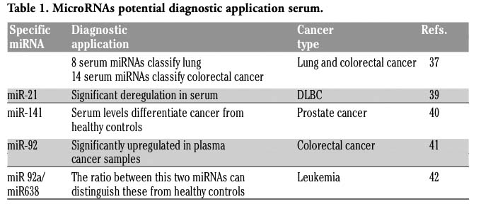
Furthermore, the search for non-invasive sensitive markers for tumor diagnosis is currently one of the most rapidly growing areas in cancer research. Very recently, miRNAs usefulness as diagnostic markers has been further expanded by studies performed in human plasma or serum.37,38 Table 1 summarizes MiRNAs detectable in serum that are diagnostically relevant.
The first serum-miRNA biomarker was reported by Lawrie et al. that showed that sera levels of miR-21 are specific for diffuse large B-cell lymphoma (DLBCL) and are associated with relapse-free survival.39
Mitchell et al. showed that serum levels of miR-141 might distinguish patients with prostate cancer from healthy controls.40
MiRNAs expression profiles from serum showed significant differences especially in lung and colorectal cancers.
Chen et al. showed that there are some "common" tumor-related miRNAs in the serum from lung and colorectal cancer patients. However, among them they identify a unique expression profile of 8 serum miRNAs in lung cancer patients, that included specific miRNAs such as miR-25 and miR-223 previously reported to be involved in tumor formation; and 14 serum miRNAs, including miR-485-5p, miR-361-3p, miR-326, miR-487b, in colorectal cancer patients.37
More recently, Ng et al. showed that miR-92 was significantly upregulated in plasma samples from colorectal cancer patients and may represent a significative marker for this type of cancer.41 The ratio of miR-92a/miR-638 in plasma has been shown to be useful for distinguishing leukemia patients from healthy individuals.42
However, although these findings reveal great impact representing a promising non-invasive method in the clinical practice for early cancer diagnosis, further independent studies on a larger cohort of patients will be necessary before any such markers can be proven to be useful in a clinical setting.
|
Table 1. MicroRNAs potential diagnostic application serum. |
Metastatic cancer of unknown primary origin accounts for 3-5% of all new cancer cases and is usually a very aggressive disease with poor prognosis.43 Clinical management of these tumors shows a high degree of variation and although many protocols have been evaluated, they show relatively little benefit, suggesting that a more accurate definition of this lesion is necessary for their treatment.
The development of technologies to study the expression levels of hundreds of miRNAs at one time demonstrated that miRNAs are an invaluable tool to distinguish tumors from normal tissue, indicating miRNAs as highly tissues specific biomarkers, as summarized in Table 2.
Two earlier studies reported that unlike with a mRNA detection method, only a modest number of miRNAs might be sufficient to classify accurately human cancers according to their developmental lineage and differentiation state.11,13 Lu et al. showed that 129 of 217 miRNAs analyzed are downmodulated in cancer compared with normal tissues, demonstrating that an miRNA-based classifier can establish the correct diagnosis of poorly differentiated cancers with far greater accuracy than an mRNA based classifier.13
In the last years numerous groups focused on identifying distinctive cancer-specific miRNA signatures that improve molecular classification of cancer.
Rosenfeld et al. have demonstrated that miRNAs can accurately identify the tissue of origin for a given cancer. They measured miRNA expression levels from 22 different tumor tissues and metastases and constructed a classifier based on 48 miRNAs with an accuracy of 90%. These results showed that a restricted number of miRNAs are sufficient to classify a cancer tissue and correlates more accurately with cancer-stage and clinicopathological features.44
Baffa et al. performed a microarray miRNA analysis on 43 matched primary tumors and corresponding lymph node metastases. This analysis identified 32 miRNAs that were differentially expressed between the 43 primary tumors and the related metastatic samples. Among them, they identified miRNAs related with the processes of tumor invasion and metastasis such as miR-200 family, miR-205 and miR 10b.45
Recently, Nass et al. showed that miR MiR-92b and miR-9/9* are specifically expressed in brain primary tumors and can be used to differentiate primary from metastatic tumors.46
These works significantly highlight that the use of miRNAs profiling outperformed mRNA in the classification of unknown primary tumor.
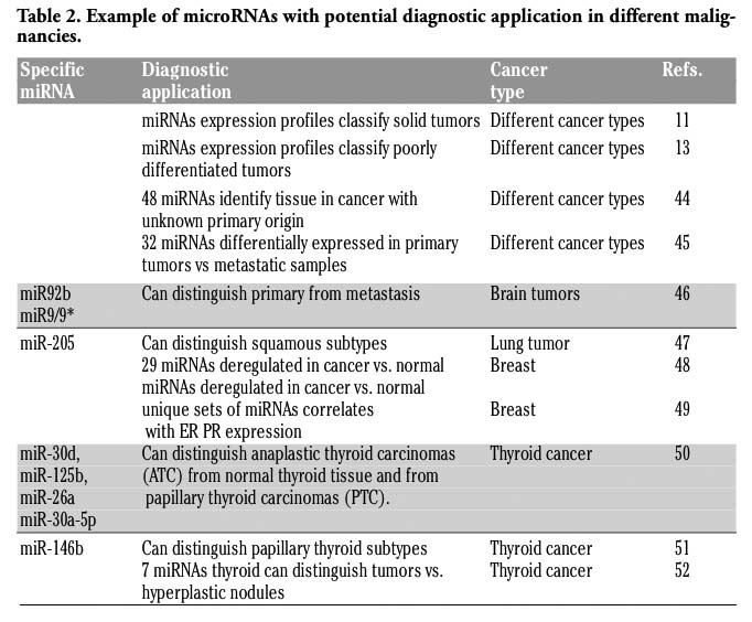 |
Table 2. Example of microRNAs with potential diagnostic application in different malignancies. |
A further aspect where miRNAs may play an advantageous role is the classification of a tumor subtype, instrumental for an appropriate treatment. The unique patterns of aberrant miRNAs expression in a specific type of cancer may improve this classification.
For example, a number of classifiers have been developed for human lung cancer and miRNAs such as miR21 and miR-205, that are located in a chromosomic region frequently amplified in this type of tumor was overexpressed.53
It has been demonstrated that miRNAs have high specificity to distinguish between non-small cell lung cancer (NSCLC), small cell lung carcinoma (SCLC), adenocarcinoma and squamous cell carcinoma.
Very recently, Lebanony showed that unlike the current diagnostic tools available, miR-205 may be highly specific to distinguish squamous from non-squamous NSCLC.47
Iorio et al. identified 29 miRNAs that may potentially act as a diagnostic molecular marker in breast cancer.48 A recent study found that a number of miRNAs were differentially expressed in breast tumor versus normal breast tissue and that unique sets of miRNA expression correlated with their ErbB2 (let-7f, let-7g, miR-107, mir-10b, miR-126, miR-154, miR-195) or ER/PR status (miR-142-5p, miR-200a, miR-205 and miR-25).49
Several studies, reviewed recently by Pallante, explored the utility of miRNA profiling for the pre-operative diagnosis of thyroid nodules.54
Particularly, Visone et al. identified a significant decrease in miR-30d, miR-125b, miR-26a and miR-30a-5p in anaplastic thyroid carcinomas (ATC) in comparison to normal thyroid tissue and from papillary thyroid carcinomas (PTC).50
Chen et al. indicated miR-146b as a potential marker to improve diagnosis of papillary thyroid carcinoma.51
Recently, Nikiforova et al. identified a set of seven miRNAs (miR-187, miR-221, miR-222, miR-146b, miR-155, miR-224, and miR-197) that were most differentially over-expressed in thyroid tumors versus hyperplastic nodules, showing high accuracy of thyroid cancer detection.52
In the past few years, there has been much evidence indicating that the miRNAs play an important role in cancer.
It has been demonstrated that there are miRNAs involved directly in cancer development controlling different cellular pathways, including cell growth, cell differentiation or apoptosis; and miRNAs that are involved indirectly in cancer development acting by targeting oncogenes and/or tumor suppressor genes.
Curently, miRNA profiling is considered the main method of investigation because it is able to show miRNAs that are differentially expressed in certain cancer tissues as compared to normal adjacent tissues and these differences may be used as biomarkers for a specific tumor.13 Moreover, an increasing number of studies highlighted significant correlations between miRNAs expression with tumor subtypes or clinical parameters. The usefulness of miRNAs in sensitively identifying the tissue of origin of metastatic cancer is a significant advance in clinical practice.
It has been demonstrated that each miRNAs may down-regulate a large number of target mRNAs, therefore the role of each miRNA may depend on the cellular context and on the pathway that they impair.
A further elucidation of the miRNA function may represent a new prospective for the identification of biomarkers in cancer.
However most of the published studies have been conducted in small and limited patient populations. To ensure the introduction of miRNAs in clinical practice as diagnostic molecular markers it will be necessary to perform further validation in multiple cohorts.
1. Griffiths-Jones S, Frocock RJ, van Dongen S, et al. MiRBase: microRNA sequences, targets and gene nomenclature. Nucleid Acids Res 2006; 34, D140-D144. [PubMed]
2. Pasquinelli AE, Reinhart BJ, Slack F, et al. Conservation of the sequence and temporal expression of let-7 heterochronic regulatory RNA. Nature 2000;408:86-9. [PubMed]
3. Floyd SK, Bowman JL. Gene regulation: ancient microRNA target sequences in plants. Nature 2004;428:485-6. [PubMed]
4. Harfe BD. MicroRNAs in vertebrate development. Curr Opin Genet Dev 2005;15:410-5. [PubMed]
5. Lau NC, Lim LP, Weinstein EG, Bartel DP. An abundant class of tiny RNAs with probable regulatory roles in Caenorhabditis elegans. Science 2001;294:858-62. [PubMed]
6. Carleton M, Cleary MA, Linsley PS. Micro- RNAs and cell cycle regulation. Cell Cycle 2007;6:2127-32. [PubMed]
7. Poy MN, Eliasson L, Krutzfeldt J, et al. A pancreatic islet-specific microRNA regulates insulin secretion. Nature 2004;432:226-30. [PubMed]
8. Landthaler M, Yalcin A, Tuschl T. The human DiGeorge syndrome critical region gene 8 and Its D. melanogaster homolog are required for miRNA biogenesis. Curr Biol 2004;14:2162-7. [PubMed]
9. Jin P, Alisch RS, Warren ST. RNA and microRNAs in fragile X mental retardation. Nat Cell Biol 2004;6:1048-53. [PubMed]
10. Meltzer PS. Cancer genomics: small RNAs with big impacts. Nature 2005;435:745-6. [PubMed]
11. Volinia S, Calin GA, Liu CG, et al. A micro- RNA expression signature of human solid tumors defines cancer gene targets. Proc Natl Acad Sci USA 2006;103:2257-61. [PubMed]
12. Lim LP, Lau NC, Garrett-Engele P, et al. Microarray analysis shows that some microRNAs downregulate large numbers of target mRNAs. Nature 2005;433:769-73. [PubMed]
13. Lu J, Getz G, Miska EA, et al. MicroRNA expression profiles classify human cancers. Nature 2005;435:834-8. [PubMed]
14. Calin GA, Sevignani C, Dumitru CD, et al. Human microRNA genes are frequently located at fragile sites and genomic regions involved in cancers. Proc Natl Acad Sci USA 2004;101:2999-3004. [PubMed]
15. Calin GA, Croce CM. MicroRNA-cancer connection: the beginning of a new tale Cancer Res 2006;66:7390-4. [PubMed]
16. Bartel DP. MicroRNAs: genomics, biogenesis, mechanism, and function. Cell 2004; 116:281-97. [PubMed]
17. Lee Y, Kim M, Han J, et al. MicroRNA genes are transcribed by RNA polymerase II. EMBO J 2004;23:4051-60. [PubMed]
18. Du T, Zamore PD. MicroPrimer: the biogenesis and function of microRNA. Development 2005;132:4645-52. [PubMed]
19. Lee Y, Ahn C, Han J, et al. The nuclear RNase III Drosha initiates microRNA processing. Nature 2003;425:415-9. [PubMed]
20. Gregory RI, Yan KP, Amuthan G, et al. The Microprocessor complex mediates the genesis of microRNAs. Nature 2004; 432:235-40. [PubMed]
21. Lund E, Güttinger S, Calado A, et al. Nuclear export of microRNA precursors. Science 2004;303:95-8. [PubMed]
22. Yi R, Qin Y, Macara IG, Cullen BR. Exportin-5 mediates the nuclear export of pre-microRNAs and short hairpin RNAs. Genes Dev 2003;17:3011-6. [PubMed]
23. Bohnsack MT, Czaplinski K, Gorlich D. Exportin 5 is a RanGTP-dependent dsRNA-binding protein that mediates nuclear export of pre-miRNAs. RNA 2004;10:185-91. [PubMed]
24. Hutvágner G, McLachlan J, Pasquinelli AE, et al. A cellular function for the RNA-interference enzyme Dicer in the maturation of the let-7 small temporal RNA. Science 2001;293:834-8. [PubMed]
25. Ketting RF, Fischer SE, Bernstein E, et al. Dicer functions in RNA interference and in synthesis of small RNA involved in developmental timing in C. elegans. Genes Dev 2001;15:2654-9. [PubMed]
26. Chendrimada TP, Gregory RI, Kumaraswamy E, et al. TRBP recruits the Dicer complex to Ago 2 for microRNA processing and gene silencing. Nature 2005;436:740-4. [PubMed]
27. Gregory RI, Chendrimada TP, Cooch N, Shiekhattar R. Human RISC couples microRNA biogenesis and posttranscriptional gene silencing. Cell 2005;123:631-40. [PubMed]
28. Reinhart BJ, Weinstein EG, Rhoades MW, et al. MicroRNAs in plants. Genes Dev 2002;16:1616-26. Erratum in: Genes Dev 2002;16:2313. [PubMed]
29. Hutvágner G, Zamore PD. A microRNA in a multiple-turnover RNAi enzyme complex. Science 2002;297:2056-60. [PubMed]
30. Deng S Calin GA Croce CM, et al. Mechanism of microRNA deregulation in human cancer. Cell Cycle 2008;7:2643-6. [PubMed]
31. O'Donnell KA, Wentzel EA, Zeller KI, et al. c-Myc-regulated microRNAs modulate E2F1 expression. Nature 2005; 435:839-43. [PubMed]
32. He L, He X, Lim LP, et al. A microRNA component of the p53 tumor suppressor network. Nature 2007; 447:1130-34. [PubMed]
33. Waldman SA, Terzic. Translating Micro RNA discovery into clinical biomarkers in cancer. JAMA 2007;297:1923-5. [PubMed]
34. Nelson PT, Baldwin DA, Scearce LM, et al. Microarray-based, high-throughput gene expression profiling of microRNAs. Nat Methods 2004;1:155-61. [PubMed]
35. Xi Y, Nakajima G, Gavin E, et al. Systematic analysis of microRNA expression of RNA extracted from fresh frozen and formalin-fixed paraffin-embedded samples. RNA 2007;13:1668-74. [PubMed]
36. Li J, Smyth P, Flavin R, et al. Comparison of miRNA expression patterns using total RNA extracted from matched samples of formalin-fixed paraffin-embedded (FFPE) cells and snap frozen cells. BMC Biotechnol 2007;7:36. [PubMed]
37. Chen X, Ba Y, Ma L, et al. Characterization of microRNAs in serum: a novel class of biomarkers for diagnosis of cancer and other diseases. Cell Res 2008;18:997-1006. [PubMed]
38. Gilad S, Meiri E, Yogev Y, et al. Serum microRNAs are promising novel biomarkers. PLoS One 2008;3:e3148. [PubMed]
39. Lawrie CH, Gal S, Dunlop HM, et al. Detection of elevated levels of tumour-associated microRNAs in serum of patients with diffuse large B-cell lymphoma. Br J Haematol 2008;141:672-5. [PubMed]
40. Mitchell PS, Parkin RK, Kroh EM, et al. Circulating microRNAs as stable blood-based markers for cancer detection. Proc Natl Acad Sci USA 2008;105:10513-8. [PubMed]
41. Ng EK, Chong WW, Jin H, et al. Differential expression of microRNAs in plasma of patients with colorectal cancer: a potential marker for colorectal cancer screening. Gut 2009;58:1375-81. [PubMed]
42. Tanaka M, Oikawa K, Takanashi M, et al. Down-regulation of miR-92 in human plasma is a novel marker for acute leukemia patients. PLoS One 2009;4:e5532. [PubMed]
43. Pimiento JM, Teso D, Malkan A, et al. Cancer of unknown primary origin: a decade of experience in a community-based hospital. Am J Surg 2007;194:833-7. [PubMed]
44. Rosenfeld N, Aharonov R, Meiri E, et al. MicroRNAs accurately identify cancer tissue origin. Nat Biotechnol 2008;26:462-9. [PubMed]
45. Baffa R, Fassan M, Volinia S, et al. MicroRNA expression profiling of human metastatic cancers identifies cancer gene targets. J Pathol 2009;219:214-21. [PubMed]
46. Nass D, Rosenwald S, Meiri E, et al. MiR-92b and miR-9/9* are specifically expressed in brain primary tumors and can be used to differentiate primary from metastatic brain tumors. Brain Pathol 2009;19:375-83. [PubMed]
47. Lebanony D, Benjamin H, Gilad S, et al. Diagnostic assay based on hsa-miR-205 expression distinguishes squamous from nonsquamous non-small-cell lung carcinoma. J Clin Oncol 2009;27:2030-7. [PubMed]
48. Iorio MV, Ferracin M, Liu CG, et al. MicroRNA gene expression deregulation in human breast cancer. Cancer Res 2005;65:7065-70. [PubMed]
49. Mattie MD, Benz CC, Bowers J, et al. Optimized high-throughput microRNA expression profiling provides novel biomarker assessment of clinical prostate and breast cancer biopsies. Mol Cancer 2006;5:24. [PubMed]
50. Visone R, Pallante P, Vecchione A, et al. Specific microRNAs are downregulated in human anaplastic carcinomas. Oncogene 2007;26:7569-95. [PubMed]
51. Chen YT, Kitabayashi N, Zhou XK, et al. MicroRNA analysis as a potential diagnostic tool for papillary thyroid carcinoma. Mod Pathol 2008;21:1139-46 [PubMed]
52. Nikiforova MN, Tseng GC, Steward D, et al. MicroRNA expression profiling of thyroid tuomrs: biological significance and diagnostic utility. J Clin Endocrinol Metab 2008;93:1600-8. [PubMed]
53. Yanaihara N, Caplen N, Bowman E, et al. Unique microRNA molecular profiles in lung cancer diagnosis and prognosis. Cancer Cell 2006;9:189-98. [PubMed]
54. Pallante L, Visone R, Croc CM, Fusco A. Deregulation of microRNA expression in follicular cell-derived human thyroid carcinomas. Endocrine-Related Cancer 2010;17 F91-F104. [PubMed]
[TOP]