
A rare ossifying trichilemmal cyst in a young female patient: case report and literature review
HTML: 10
All claims expressed in this article are solely those of the authors and do not necessarily represent those of their affiliated organizations, or those of the publisher, the editors and the reviewers. Any product that may be evaluated in this article or claim that may be made by its manufacturer is not guaranteed or endorsed by the publisher.
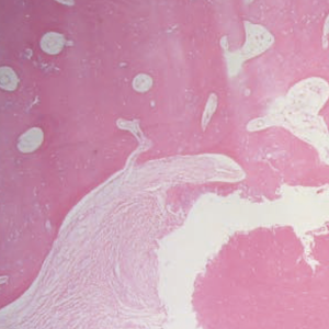
Trichilemmal cysts (TCs) constitute the second most common cutaneous cysts and are mostly presented on the scalp of middleaged women. Therefore, it is unusual for a young person to have a TC and it is extremely rare for a TC to be ossified. In the literature, only 8 cases of TCs with concomitant ossification have been described. We report the case of a 22-year-old female who presented with a scalp nodule and was treated via surgical excision of the lesion. The pathology examination of the surgical specimen revealed a lesion consisting of a multilayered squamous epithelium of slightly eosinophilic maturing keratinocytes. There was no granular layer, whereas the core of the lesion was occupied by mature bone tissue with calcium deposits. The definite diagnosis of the pathology report was ossifying TC. The aim of this report is, to enlighten clinicians about this rare pathological entity.
Pusiol T, Morichetti D, Zorzi MG, Francesco P. Ossifying trichilemmal cyst. Am J Dermatopathol 2011;33:867-8.
Kamyab K, Kianfar N, Dasdar S, et al. Cutaneous cysts: a clinicopathologic analysis of 2,438 cases. Int J Dermatol 2020;59:457-62.
Kolodney MS, Coman GC, Smolkin MB, et al. Hereditary trichilemmal cysts are caused by two hits to the same copy of the phospholipase c delta 1 gene (PLCD1). Sci Rep 2020;10:6035.
Hörer S, Marrakchi S, Radner FP, et al. A monoallelic two-hit mechanism in PLCD1 explains the genetic pathogenesis of hereditary trichilemmal cyst formation. J Invest Dermatol 2019;139:2154-63.
Baldovini C, Rosini F, Marucci G. Osseous metaplasia and mature bone formation with extramedullary hematopoiesis in trichilemmal cyst. Am J Dermatopathol 2014;36:444-6.
Fletcher CD. Calcifying and ossifying soft tissue lesions presenting in the skin. J Cutan Pathol 1996;23:297.
Civatte J, Tsoïtis G, Le Roux P. Perforating ossified (trichilemmal) "sebaceous" cyst. Apropos of a case. Ann Dermatol Syphiligr (Paris) 1974;101:155-70.
Mommers XA, Henault B, Aubriot MH, et al. Multiple ossifying trichilemmal cysts of the scalp: a familial case. Rev Stomatol Chir Maxillofac 2012;113:53-6.
Bär M, Kayser DM, Trniková M, Grunow N. Perforating and ruptured trichilemmal cyst with metaplastic ossification. Pathologe 2012;33:459-62.
Bulut AŞ. Trichilemmal cyst with ossification and marrow formation: a case report. J Turk Acad Dermatol 2012;6:1263c4.
Abreu Velez AM, Brown VM, Howard MS. An inflamed trichilemmal (pilar) cyst: not so simple? N Am J Med Sci 2011;3:431-4.
He P, Chen W, Zhang Q, et al. Distinguishing a trichilemmal cyst from a pilomatricoma with ultrasound. J Ultrasound Med 2020;39:1939-45.
Singh P, Usman A, Motta L, Khan I. Malignant proliferating trichilemmal tumour. BMJ Case Rep 2018;2018:bcr2018224460.
Osto M, Parry N, Rehman R, et al. Malignant proliferating trichilemmal tumor of the scalp: a systematic review. Am J Dermatopathol 2021;43:851-66.
Anolik R, Firoz B, Walters RF, et al. Proliferating trichilemmal cyst with focal calcification. Dermatol Online J 2008;14:25.
Copyright (c) 2022 the Author(s)

This work is licensed under a Creative Commons Attribution-NonCommercial 4.0 International License.
PAGEPress has chosen to apply the Creative Commons Attribution NonCommercial 4.0 International License (CC BY-NC 4.0) to all manuscripts to be published.
Downloads
Citations


