
Environmental factors in autoimmune bullous diseases with a focus on seasonality: new insights
Accepted: 8 February 2023
HTML: 10
All claims expressed in this article are solely those of the authors and do not necessarily represent those of their affiliated organizations, or those of the publisher, the editors and the reviewers. Any product that may be evaluated in this article or claim that may be made by its manufacturer is not guaranteed or endorsed by the publisher.
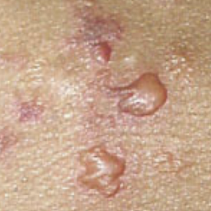
Autoimmune bullous diseases are a heterogeneous group of rare conditions clinically characterized by the presence of blisters and/or erosions on the skin and the mucous membranes. Practically, they can be divided into two large groups: the pemphigoid group and the pemphigus group, depending on the depth of the autoimmune process on the skin. A family history of autoimmune diseases can often be found, demonstrating that genetic predisposition is crucial for their development. Moreover, numerous environmental risk factors, such as solar radiation, drugs, and infections, are known. This study aimed to evaluate how seasonality can affect the trend of bullous pemphigoid and pemphigus vulgaris, especially considering the number of hospitalizations recorded over the course of individual months. The total number of hospitalizations in the twelve months of the year was evaluated. Moreover, blood chemistry assay and, for some patients, enzyme-linked immunosorbent assay were executed to evaluate antibodies. Regarding the severity of the disease, the bullous pemphigoid area index and the pemphigus disease area index score systems were used. Results showed a complex interplay between environmental factors such as seasons and autoimmune conditions.
Didona D et al. Humoral Epitope Spreading in Autoimmune Bullous Diseases. Front Immunol. 2018 Apr 17;9:779. DOI: https://doi.org/10.3389/fimmu.2018.00779
Wang WM et al. Role of B cells in immune-mediated dermatoses. Mol Immunol. 2020 Oct;126:95-100. DOI: https://doi.org/10.1016/j.molimm.2020.07.016
Pile HD et al. Drug Induced Pemphigus. StatPearls Publishing; 2020 Jan. 2020 Aug 25.
Hibi A et al. Dipeptidyl peptidase-4 inhibitor-associated bullous pemphigoid, likely triggered by scabies, in a hemodialysis patient with human leukocyte antigen-DQB1*03:01. CEN Case Rep. 2020 Aug; 9(3): 189–194. DOI: https://doi.org/10.1007/s13730-020-00452-2
Tur E et al. Contributing exogenous factors in pemphigus. Int J Dermatol. 1997 Dec;36(12):888-93. DOI: https://doi.org/10.1046/j.1365-4362.1997.00334.x
Krain LS (1974) Pemphigus. Epidemiologic and survival characteristics of 59 patients, 1955–1973. Arch Dermatol 110:862–865. DOI: https://doi.org/10.1001/archderm.110.6.862
Ruocco E, Ruocco V, Lo Schiavo A, Brunetti G, Wolf R (2014) Viruses and pemphigus: an intriguing never-ending story. Dermatology 229:310–315. DOI: https://doi.org/10.1159/000365845
Mohammadi F et al. The potential roles of herpesvirus and cytomegalovirus in the exacerbation of pemphigus vulgaris. Dermatol Pract Concept. 2018 Oct 31;8(4):262-271. DOI: https://doi.org/10.5826/dpc.0804a03
Ren Z et al. Association between climate, pollution and hospitalization for pemphigus in the USA. Clin Exp Dermatol. 2019 Mar;44(2):135-143. DOI: https://doi.org/10.1111/ced.13650
Fisher KR et al. Pesticide-associated pemphigus vulgaris. Cutis 2008 Jul;82(1):51-4.
Tsankov N et al. Epidemiology of pemphigus in Sofia, Bulgaria. A 16-year retrospective study (1980-1995). Int J Dermatol. 2000 Feb;39(2):104-8. DOI: https://doi.org/10.1046/j.1365-4362.2000.00864.x
Kyriakis KP et al. Environmental factors influencing the biologic behavior of patterns of pemphigus vulgaris: epidemiologic approach. Int J Dermatol. 1995 Mar;34(3):181-5. DOI: https://doi.org/10.1111/j.1365-4362.1995.tb01563.x
Salmanpour R et al. Epidemiology of pemphigus in south-western Iran: A 10-year retrospective study (1991–2000). Int J Dermatol. 2006 Feb;45(2):103-5. DOI: https://doi.org/10.1111/j.1365-4632.2004.02374.x
Robati RM et al. Pemphigus vulgaris and season: are they really related or not? J Eur Acad Dermatol Venereol. 2011 Oct;25(10):1235-6. DOI: https://doi.org/10.1111/j.1468-3083.2010.03867.x
Marzano AV et al. Evidence for vitamin D deficiency and increased prevalence of fractures in autoimmune bullous skin diseases. Br J Dermatol. 2012 Sep;167(3):688-91. DOI: https://doi.org/10.1111/j.1365-2133.2012.10982.x
Marzano AV et al. Vitamin D and skeletal health in autoimmune bullous skin diseases: A case control study. Orphanet Journal of Rare Diseases, 10 (2015), p. 8. DOI: https://doi.org/10.1186/s13023-015-0230-0
Tukaj S et al. Vitamin D status in bullous pemphigoid patients. The British Journal of Dermatology, 168 (2013), pp. 873-874. DOI: https://doi.org/10.1111/bjd.12037
Zarei M et al. Evaluation of Vitamin D Status in Newly Diagnosed Pemphigus Vulgaris Patients. Iran J Public Health. 2014 Nov; 43(11): 1544–1549.
Tavalkopour S et al. Pemphigus trigger factors: special focus on pemphigus vulgaris and pemphigus foliaceus. Arch Dermatol Res (2018) 310:95–106. DOI: https://doi.org/10.1007/s00403-017-1790-8
Shear NH. Diagnosing cutaneous adverse reactions to drugs. Archives of Dermatology. 1990;126(1):94–97. DOI: https://doi.org/10.1001/archderm.126.1.94
Brenner S et al. Drug-induced pemphigus. Clin Dermatol. Jul-Aug 2011;29(4):455-7. DOI: https://doi.org/10.1016/j.clindermatol.2011.01.016
Lo Schiavo A et al. Bullous pemphigoid: Etiology, pathogenesis, and inducing factors: Facts and controversies. Clinics in Dermatology (2013)31, 391–399. DOI: https://doi.org/10.1016/j.clindermatol.2013.01.006
Fang H et al. Association of HLA class I and class II alleles with bullous pemphigoid in Chinese Hans. J Dermatol Sci. 2018 Mar;89(3):258-262. DOI: https://doi.org/10.1016/j.jdermsci.2017.11.014
Ujiie H et al. HLA-DQB1*03:01 as a Biomarker for Genetic Susceptibility to Bullous Pemphigoid Induced by DPP-4 Inhibitors. J Invest Dermatol. 2018 May;138(5):1201-1204. DOI: https://doi.org/10.1016/j.jid.2017.11.023
Moro F et al. Bullous Pemphigoid: Trigger and Predisposing Factors. Biomolecules. 2020 Oct; 10(10): 1432. DOI: https://doi.org/10.3390/biom10101432
Halevy S, Cohen AD, Grossman N. Clinical implications of in vitro drug-induced interferon gamma release from peripheral blood lymphocytes in cutaneous adverse drug reactions. Journal of the American Academy of Dermatology. 2005;52(2):254–261. DOI: https://doi.org/10.1016/j.jaad.2004.05.006
De Simone C et al. Exacerbation of pemphigus after influenza vaccination. Clin Exp Dermatol. 2008 Nov;33(6):718-20. DOI: https://doi.org/10.1111/j.1365-2230.2008.02835.x
Kano Y et al. Pemphigus foliaceus induced by exposure to sunlight. Report of a case and analysis of photochallenge-induced lesions. Dermatology. 2000;201(2):132-8. DOI: https://doi.org/10.1159/000018456
Charoenngam N et al. Immunologic Effects of Vitamin D on Human Health and Disease. Nutrients. 2020 Jul 15;12(7):2097. DOI: https://doi.org/10.3390/nu12072097
Sarre ME et al. Association between bullous pemphigoid and hypovitaminosis D in older inpatients: Results from a case-control study. Eur J Intern Med. 2016 Jun;31:25-8. DOI: https://doi.org/10.1016/j.ejim.2016.02.004
Copyright (c) 2023 the Author(s)

This work is licensed under a Creative Commons Attribution-NonCommercial 4.0 International License.
PAGEPress has chosen to apply the Creative Commons Attribution NonCommercial 4.0 International License (CC BY-NC 4.0) to all manuscripts to be published.
Downloads
Citations


