
Mohs micrographic surgery: the experience of the Dermatology Unit of the University of Milan confirms the superiority over traditional surgery in high-risk non-melanoma skin cancers
HTML: 6
All claims expressed in this article are solely those of the authors and do not necessarily represent those of their affiliated organizations, or those of the publisher, the editors and the reviewers. Any product that may be evaluated in this article or claim that may be made by its manufacturer is not guaranteed or endorsed by the publisher.
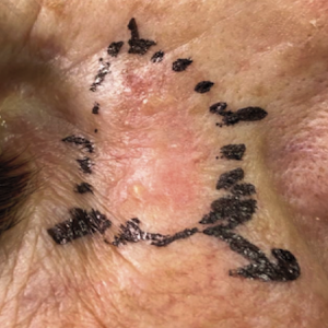
The constant increase in the incidence of non-melanoma skin cancers (NMSC) makes their treatment a topic of paramount interest. Because most NMSC tend to develop in visible areas such as the head-neck area, it is a priority to choose the less destructive therapy and more appropriate reconstructive technique. Mohs Micrographic Surgery (MMS) represents the treatment of choice for skin tumors in critical sites, recurrent tumors and tumors with aggressive histologic features. We collected patients affected by NMSC who underwent MMS at the Dermatology Unit of IRCCS Fondazione Ca' Granda, Milan, in the period March 2017-December 2021. One hundred and fifty-nine patients were enrolled in this retrospective observational study. The excision margins were chosen based on a dermoscopic evaluation. The main histological diagnoses were basal cell carcinoma (145, 91.2%) and squamous cell carcinoma (10, 6.3%), in areas with high functional or anatomical value. 121 out of 159 surgeries did not require further enlargement after the removal of the clinically and dermoscopically visible lesion, but in 38 cases (23.9% of cases) the pathologist required at least one subsequent enlargement, due to the persistence of neoplasm at the bottom or at the margins of the lesion. Only one recurrence has been reported so far. MMS is a pathology-controlled surgery with high intrinsic value because of the low risk of recurrences and should be routinely adopted for high-risk NMSC.
Cohen DK, Goldberg DJ. Mohs micrographic surgery: past, present, and future. Dermatol Surg 2019;45:329-39.
Lewis KG, Weinstock MA. Non melanoma skin cancer mortality (1988–2000): the rhode island follow-back study. Arch Dermatol 2004;140:837–42.
Rogers HW, Weinstock MA, Feldman SR, Coldiron BM. Incidence estimated of non melanoma skin cancer (keratinocytes carcinomas) in the US population, 2012. JAMA Dermatol 2015;151:1081-6.
Langtry J.A.A. International perspective of mohs micrographic surgery: Europe. In: Nouri K, eds. Mohs Micrographic Surgery. London, UK: Springer; 2012.
Peris K, Fargnoli MC, Garbe C, et al. Diagnosis and treatment of basal cell carcinoma: European consensus-based interdisciplinary guideline. Eur J Cancer 2019;118:10-34.
Alexander J. Stratigos, Claus Garbe, Clio Dessinioti, et al. European interdisciplinary guideline on invasive squamous cell carcinoma of the skin: part 2. Treatment. Eur J Cancer 2020;128:83-102.
Ad Hoc Task Force, Connolly SM, Baker DR, et al. AAD/ACMS/ASDSA/ASMS 2012 appropriate use criteria for Mohs micrographic surgery: a report of the American Academy of Dermatology, American College of Mohs Surgery, American Society for Dermatologic Surgery Association, and the American Society for Mohs Surgery. J Am Acad Dermatol 2012;67:531-50.
Work Group; Invited Reviewers, Kim JYS, et al. Guidelines of care for the management of cutaneous squamous cell carcinoma. J Am Acad Dermatol 2018;78:560–78.
Work Group; Invited Reviewers, Kim JYS, et al. Guidelines of care for the management of basal cell carcinoma. J Am Acad Dermatol 2018;78:540–59.
Sharma AN, Foulad DP, Doan L, et al. Mohs surgery for the treatment of lentigo maligna and lentigo maligna melanoma - a systematic review. J Dermatolog Treat 2021;32:157-63.
Rodríguez-Jiménez P, Jimenez YD, Reolid A, et al. State of the art of Mohs surgery for rare cutaneous tumors in the Spanish registry of Mohs surgery (REGESMOHS). Int J Dermatol 2020;59:321–5.
Lance D. Wood MD. An overview of mohs micrographic surgery for the treatment of basal cell carcinoma. Dermatol Clin 2011;29:153-60.
Gladstone HB, Stewart D. An algorithm for the reconstruction of complex facial defects. Skin Therapy Lett 2007;12:6–9.
Cerci F, Kubo EM, Werner B, Tolkachjov SN. Surgical margins required for basal cell carcinomas treated with Mohs micrographic surgery according to tumor features. J Am Acad Dermatol 2020;83:493-500.
Alam M, Berg D, Bhatia A, et al. Association between number of stages in Mohs micro-graphic surgery and surgeon-, patient-, and tumor-specific features: a cross-sectional study of practice patterns of 20 early- and mid-career Mohs surgeons. Dermatol Surg 2010;36:1915-20.
Alam M, Helenowksi IB, Cohen JL, et al. Association between type of reconstruction after Mohs micrographic surgery and surgeon-, patient-, and tumor-specific features: a cross-sectional study. Dermatol Surg 2013;39:51–5.
Cook J, Zitelli JA. Mohs micrographic surgery: a cost analysis. J Am Acad Dermatol 1998;39:698–703.
Alsaif A, Hayre A, Karam M, et al. Mohs micrographic surgery versus standard excision for basal cell carcinoma in the head and neck: systematic review and meta-analysis. Cureus 2021;13:e19981.
Udkoff J, Beal BT, Brodland DG, Knackstedt T. Cost effectiveness of intermediate-risk squamous cell carcinoma treated with mohs micrographic surgery compared with wide local excision. J Am Acad Dermatol 2021;86:303-11.
Copyright (c) 2022 the Author(s)

This work is licensed under a Creative Commons Attribution-NonCommercial 4.0 International License.
PAGEPress has chosen to apply the Creative Commons Attribution NonCommercial 4.0 International License (CC BY-NC 4.0) to all manuscripts to be published.
Downloads
Citations


